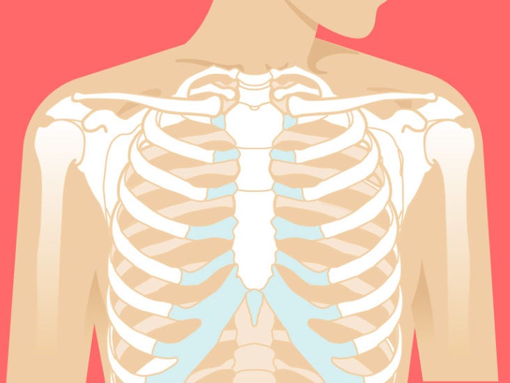
Anatomy Of The Upper Chest Area : Upper Chest Workouts Mdarr5949 / Anatomy of the chest & abdomen.
byAdmin-
0
Anatomy Of The Upper Chest Area : Upper Chest Workouts Mdarr5949 / Anatomy of the chest & abdomen.. • acromion • clavicle • deltoid ( im injections) • humerus axilla(armpit). The superior vena cava (svc) is seen in the right paratracheal area, typically representing the right superior mediastinal contour. The lungs are assessed and described by dividing them into upper, middle and lower zones. Anatomy of peritoneum and mesentery. Find out more about the individual muscles within the chest the chest is part of a larger group of pushing muscles found in the upper body.
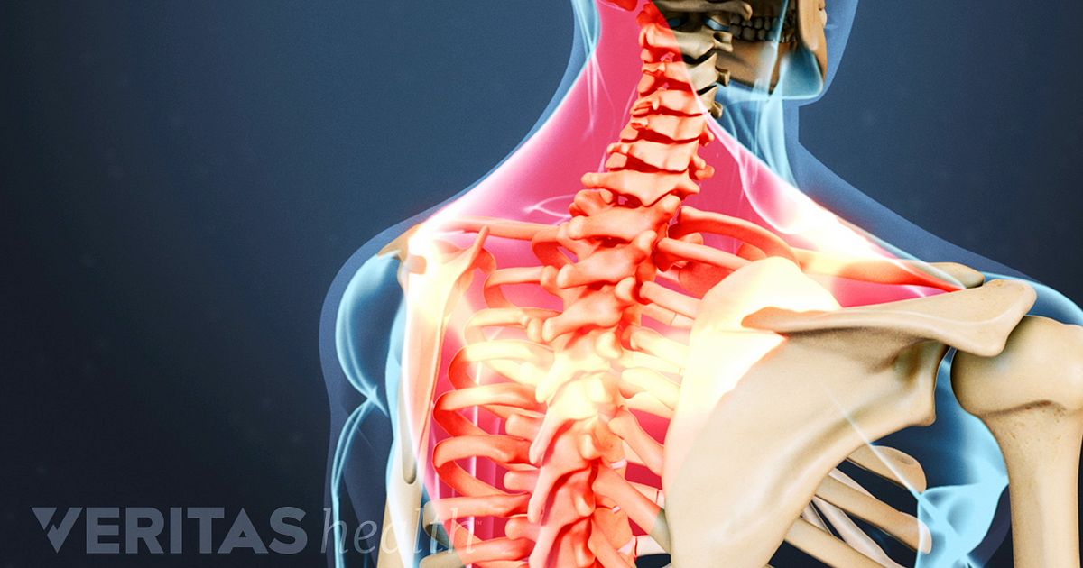
The upper chest has two main functions: • pyramidal space between the upper lateral chest and the innerside of the arm. Upper back pain and chest pain can occur together. I will therefore split the chest up into three parts: For the purpose of description the lungs are divided into zones:
Sternum Anatomy Location Function Pain Injuries from post.healthline.com The lungs are assessed and described by dividing them into upper, middle and lower zones. Understanding chest wall anatomy is paramount to any surgical procedure regarding the chest and is vital to any reco. This page provides an overview of the chest muscle group. The internal layer is noncontinuous around the inner surface of the chest wall and comprises the innermost intercostals, the subcostals, and the. We're looking at the anatomy of an upper endoscopy. It describes the theatre of events. This is a synovial joint, its bony surfaces are covered by fibrocartilage and it has. An important palpable feature on the anterior chest wall.
The upper chest has two main functions:
You can use your stethoscope to listen to the heart beat and inspect chest movements to help determine how well the patient is breathing. The upper posterior border of the heart is formed by the left atrium. Chest workouts to target different chest muscles. • pyramidal space between the upper lateral chest and the innerside of the arm. The best upper chest workout will. • acromion • clavicle • deltoid ( im injections) • humerus axilla(armpit). Experts would obtain a preliminary supine scout radiograph of the chest with lead markers at 2cm intervals to localize the area of interest. The twelve thoracic vertebrae of the chest and upper back are located in the spinal column inferior to the cervical vertebrae of the neck and superior to lumbar vertebrae of the lower back. Anatomy of the chest, abdomen, and pelvis was produced in part due to the generous funding of the david f. Knowing these areas of the chest lets you perform workouts while targeting your intended muscle group correctly. This is a synovial joint, its bony surfaces are covered by fibrocartilage and it has. Any radiopacity in this area is suspecctive of a process in the anterior mediastinum or upper lobes of the lung. Human anatomy for muscle, reproductive, and skeleton.
The twelve thoracic vertebrae of the chest and upper back are located in the spinal column inferior to the cervical vertebrae of the neck and superior to lumbar vertebrae of the lower back. The anterior of the chest is a main area for physical examination. The embryologic and anatomic basis of modern surgery. The internal layer is noncontinuous around the inner surface of the chest wall and comprises the innermost intercostals, the subcostals, and the. Anatomy is to physiology as geography is to history:
What Causes Upper Back And Chest Pain from embed.widencdn.net It describes the theatre of events. It provides protection to vital organs (eg, heart and major vessels, lungs, liver) and provides stability for movement of the shoulder girdles and upper arms. I will therefore split the chest up into three parts: Thoracic vertebrae interlock tightly by overlapping their spinous processes, giving stability to the spine in this. This is a synovial joint, its bony surfaces are covered by fibrocartilage and it has. The opening of the upper chest and thorax. Chest auscultation requires the chest and back to be exposed, so measures should be taken to this technique allows you to compare one side of the chest with the other in a systematic manner and starting with the upper lobe move to the middle lobe, and finally the lower lobe at the bottom (ferns. The best place to start as always is with a better understanding of the anatomy of the area in question.
Enlargement will result in bulging of the.
The opening of the upper chest and thorax. Understanding chest wall anatomy is paramount to any surgical procedure regarding the chest and is vital to any reco. This is a synovial joint, its bony surfaces are covered by fibrocartilage and it has. Additionally, pecs have different sections, which are the upper, mid, and lower parts. Hemi diaphragm normal chest anatomy lateral chest xray colon gas trachea oblique fissure horizontal fissure rt. Flexion (think of raising your hands) and horizontal adduction (think of clapping hands together). It describes the theatre of events. Anatomy of peritoneum and mesentery. Upper back pain and chest pain can occur together. Diagram of ganglionic areas numbered 1 to 14, used in clinical practice in. Located at the level of the intervertebral disc between t4 and t5. Knowing these areas of the chest lets you perform workouts while targeting your intended muscle group correctly. I will therefore split the chest up into three parts:
This is a synovial joint, its bony surfaces are covered by fibrocartilage and it has. All about the chest muscles function of the chest muscles. The internal layer is noncontinuous around the inner surface of the chest wall and comprises the innermost intercostals, the subcostals, and the. Enlargement will result in bulging of the. Chest auscultation requires the chest and back to be exposed, so measures should be taken to this technique allows you to compare one side of the chest with the other in a systematic manner and starting with the upper lobe move to the middle lobe, and finally the lower lobe at the bottom (ferns.
Thoracic Wall And Breast Illustrations from www.imaios.com • acromion • clavicle • deltoid ( im injections) • humerus axilla(armpit). Located at the level of the intervertebral disc between t4 and t5. Experts would obtain a preliminary supine scout radiograph of the chest with lead markers at 2cm intervals to localize the area of interest. Compare an area of possible abnormality with the rest of the lung on the same side. The superior vena cava (svc) is seen in the right paratracheal area, typically representing the right superior mediastinal contour. • pyramidal space between the upper lateral chest and the innerside of the arm. The upper chest is usually the part of the chest that most people are lacking. Swensen fund for innovation in teaching.
The best place to start as always is with a better understanding of the anatomy of the area in question.
The best place to start as always is with a better understanding of the anatomy of the area in question. We're looking at the anatomy of an upper endoscopy. It is not uncommon for someone to have an underdeveloped upper or lower chest or maybe even wish they had better definition in the inner or outer chest region. Anatomy of the chest & abdomen. Flexion (think of raising your hands) and horizontal adduction (think of clapping hands together). The opening of the upper chest and thorax. Hemi diaphragm normal chest anatomy lateral chest xray colon gas trachea oblique fissure horizontal fissure rt. Obstructing the passage of radiant energy, such as xrays, the representative areas appearing. Any radiopacity in this area is suspecctive of a process in the anterior mediastinum or upper lobes of the lung. Chest workouts to target different chest muscles. Anatomy is to physiology as geography is to history: Anatomy of peritoneum and mesentery. The upper chest has two main functions: